The pathogen
The main agent is Mycoplasma agalactiae, although Mycoplasma mycoides subsp. capri, Mycoplasma capricolum subsp. capricolum and Mycoplasma putrefaciens can also be found in goats.
How does the disease occur?
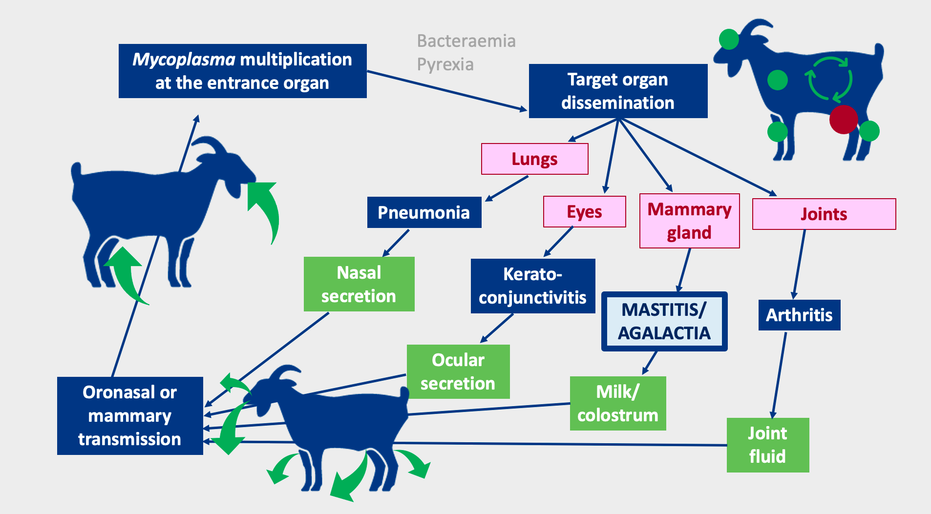
Transmission can be by oronasal route through nasal or ocular discharge or secretion from joint lesions of infected animals. Contaminated surfaces are the main source of infection, such as feeders, drinkers, or bedding. Kids or lambs can be infected through colostrum or milk.

Females are also infected through the mammary route, through teat cups or the hands of milkers, which may be contaminated.
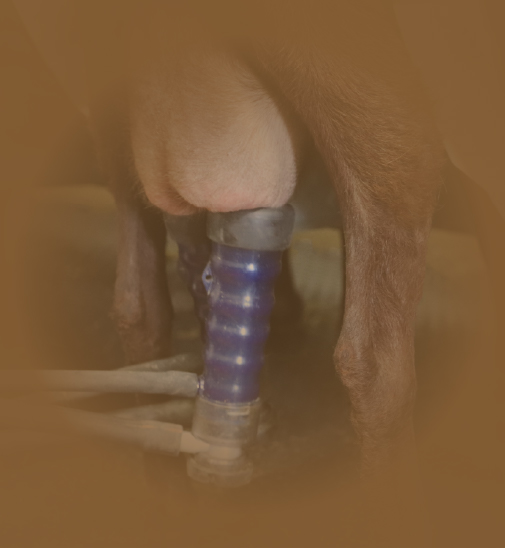
Females are mainly infected by the mammary route, through contaminated teat cups
When the animal becomes infected, the pathogen multiplies and then spreads through the target organs: mammary gland, lung, eyes, joints… Symptoms include mastitis, pneumonia, keratoconjunctivitis, arthritis, septicemia, occasional abortion and orchitis in males.
Herds can suffer outbreaks or be chronic, having a high incidence of subclinical mastitis. Chronic herds are common in goats, in which there are asymptomatic carriers.
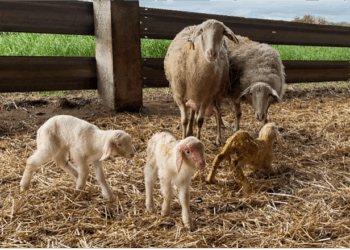
Asymptomatic carriers maintain the disease in the herd
What happens at the udder?
Initially, the udder appears swollen and hot, and later it becomes flaccid and full of connective tissue. At this stage, the milk becomes more watery, yellowish or greenish (and can become purulent) and its production is reduced.
Finally, the udder atrophies and agalactia occurs due to the necrosis of the glandular epithelia and the compression exerted by the fibrosis of the tissue.
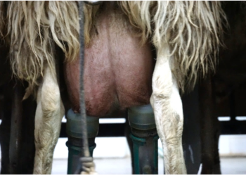
The infection causes atrophy of the udder, and therefore milk production ceases
Conclusions
● Diverse aetiology.
● Great variability of symptoms.
● Presence of asymptomatic carriers.
● Great economic cost due to the loss of dairy production.
Article written by:
Tania Perálvarez Puerta. Product Manager Small Ruminants Unit – HIPRA


